
Scientia Silvae Sinicae ›› 2021, Vol. 57 ›› Issue (11): 152-157.doi: 10.11707/j.1001-7488.20211115
Previous Articles Next Articles
Shitao Wen,Linxin Dai,Xing Liu,Jianfeng Ma*
Received:2020-09-11
Online:2021-11-25
Published:2022-01-12
Contact:
Jianfeng Ma
CLC Number:
Shitao Wen,Linxin Dai,Xing Liu,Jianfeng Ma. The Ultrastructure and Polysaccharides Composition of Middle Lamella in Rattan Cane (Calamus simplicifolius)[J]. Scientia Silvae Sinicae, 2021, 57(11): 152-157.
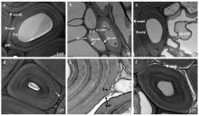
Fig.1
TEM image of cross section of rattan stem a: Control fiber; b: Parenchyma; c, d, e, f: Delignified sections of rattan cane. ff-ccml: Cell corner middle lamella among adjacent fibers; pp-ccml: Cell corner middle lamella among adjacent parenchyma; fp-ccml: Cell corner middle lamella between fiber and parenchyma; ff-cml: Compound middle lamella among adjacent fibers; pp-cml: Compound middle lamella among adjacent parenchyma; fp-cml: Compound middle lamella between fiber and parenchyma; f-s: Fiber secondary wall; p-s: Parenchyma secondary wall; pw: Primary wall. The same below."

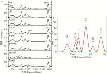
Fig.2
Average Raman spectra extracted from various cell wall layers (a) and deconvoluted spectra from fiber secondary wall (b)"

Table 1
Raman band assignments of cell wall polysaccharides and lignin"
| 波数Wavenumbers/cm-1 | 归属Assignment |
| 2 942 | 木质素及碳水化合物C—H和C—H2伸缩振动 C—H and C—H2 stretching in lignin and carbohydrate |
| 2 897 | 碳水化合物C—H和C—H2伸缩振动 C—H and C—H2 stretching in carbohydrate |
| 1 660 | 木质素芳香环共轭的松伯醇CC及松伯醛CO伸缩振动 Ring conjungated CC stretching of coniferyl alcohol; CO stretching of coniferaldehyde in lignin |
| 1 620 | 木质素芳香环共轭的松伯醛CC伸缩振动 Ring conjungated CC stretching of coniferaldehyde in lignin |
| 1 598 | 木质素芳香环伸缩振动 Aryl ring stretching symmetric in lignin |
| 1 479 | 纤维素C—H2和C—OH剪切振动 C—H2 and C—OH scissoring in cellulose |
| 1 453 | 木质素C—H3剪切振动,芳香环面外C—H3弯曲振动 C—H3 scissoring, C—H3 out-of-plane bending in lignin |
| 1 378 | 木质素酚羟基弯曲振动,C—H伸缩振动 Phenolic O—H bending, C—H stretching in lignin |
| 1 338 | 纤维素C—H2弯曲振动 C—H2 bending in cellulose |
| 1 318 | 木聚糖C—H弯曲振动 C—H bending in xylan |
| 1 247 | 果胶C—H弯曲振动 C—H bending in pectin |
| 1 122 | 纤维素糖苷键C—O—C对称伸缩振动 C—O—C symmetric stretching in cellulose |
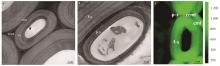
Fig.3
TEM images showing wall ultrastructure of rattan fiber (a) and parenchyma (b) as well as Raman image showing the morphology of fiber and parenchyma (c)"


Fig.4
Raman images showing the lignin (a), cellulose (b), xylan (c) and pectin (d) distribution within the fiber and parenchyma wall"

| 冯龙, 孙存举, 毕文思, 等. 毛竹薄壁细胞组分分布及取向显微成像研究. 光谱学与光谱分析, 2020, 40 (9): 307- 311. | |
| Feng L , Sun C J , Bi W S , et al. The distribution and orientation of cell wall components of moso bamboo parenchyma. Spectroscopy and Spectral Analysis, 2020, 40 (9): 2957- 2961. | |
| 刘杏娥, 金克霞, 崔贺帅, 等. 黄藤细胞壁木质素区域化学分子光谱成像研究. 光谱学与光谱分析, 2017, 37 (10): 3138- 3144. | |
| Liu X E , Jin K X , Cui H S , et al. The lignin topochemistry of Daemonorops margaritae (Hance) Becc. by molecular spectroscopic imaging. Spectroscopy and Spectral Analysis, 2017, 37 (10): 3138- 3144. | |
|
Abasolo W P , Yoshida M , Yamamoto H , et al. Microfibril angle determination of rattan fibers and its influence on the properties of the cane. Holzforschung, 2000, 54 (4): 437- 442.
doi: 10.1515/HF.2000.072 |
|
|
Agarwal U P , Atalla R H . In-situ Raman microprobe studies of plant cell walls: macromolecular organization and compositional variability in the secondary wall of Picea mariana (Mill.) B. S. P. Planta, 1986, 169 (3): 325- 332.
doi: 10.1007/BF00392127 |
|
|
Agarwal U P , Ralph S A . FT-Raman spectroscopy of wood: identifying contributions of lignin and carbohydrate polymers in the spectrum of black spruce (Picea mariana). Applied Spectroscopy, 1997, 51 (11): 1648- 1655.
doi: 10.1366/0003702971939316 |
|
|
Bhat K M , Nasser K M M , Thulasidas P K . Anatomy and identification of south Indian rattans (Calamus species). IAWA Journal, 1993, 14 (1): 63- 76.
doi: 10.1163/22941932-90000578 |
|
|
Bock P , Nousiainen P , Elder T , et al. Infrared and Raman spectra of lignin substructures: dibenzodioxocin. Journal of Raman Spectroscopy, 2020, 51 (3): 422- 431.
doi: 10.1002/jrs.5808 |
|
|
Cosgrover D J . Diffuse growth of plant cell walls. Plant Physiology, 2018, 176 (1): 16- 27.
doi: 10.1104/pp.17.01541 |
|
|
Donaldson L A . Lignification and lignin topochemistry-an ultrastructural view. Phytochemistry, 2001, 57 (6): 859- 873.
doi: 10.1016/S0031-9422(01)00049-8 |
|
|
Gibson L J . The hierarchical structure and mechanics of plant materials. Journal of the Royal Society Interface, 2012, 9 (76): 2749- 2766.
doi: 10.1098/rsif.2012.0341 |
|
| Gierlinger N , Schwanninger M . The potential of Raman microscopy and Raman imaging in plant research. Spectroscopy, 2012, 21 (2): 69- 89. | |
|
Gierlinger N . New insights into plant cell walls by vibrational microspectroscopy. Applied Spectroscopy Reviews, 2018, 53 (7): 517- 551.
doi: 10.1080/05704928.2017.1363052 |
|
|
Grünwald C , Ruel K , Kim Y S , et al. On the cytochemistry of cell wall formation in poplar trees. Plant Biology, 2002, 4 (1): 13- 21.
doi: 10.1055/s-2002-20431 |
|
|
Higuchi T . Lignin biochemistry: biosynthesis and biodegradation. Wood Science and Technology, 1990, 24 (1): 23- 63.
doi: 10.1021/es00071a002 |
|
|
Jin K X , Liu X E , Jiang Z H , et al. Delignification kinetics and selectivity in poplar cell wall with acidified sodium chlorite. Industrial Crops and Products, 2019, 136, 87- 92.
doi: 10.1016/j.indcrop.2019.04.067 |
|
|
Kačuráková M , Wellner M , Ebringerova N , et al. Characterisation of xylan-type polysaccharides and associated cell wall components by FT-IR and FT-Raman spectroscopies. Food Hydrocolloids, 1999, 13 (1): 35- 41.
doi: 10.1016/S0268-005X(98)00067-8 |
|
|
Kim J S , Awano T , Yoshinaga A , et al. Immunolocalization and structural variations of xylan in differentiating earlywood tracheid cell walls of Cryptomeria japonica. Planta, 2010, 232 (4): 817- 824.
doi: 10.1007/s00425-010-1225-7 |
|
|
Kim Y S , Lee K H , Kim J S , et al. Lignin masks the presence of fibrillar network structure in the cell corner middle lamella(CCML). Holzforschung, 2015, 69 (1): 121- 126.
doi: 10.1515/hf-2014-0032 |
|
|
Mellerowicz E J , Baucher M , Sundberg B , et al. Unravelling cell wall formation in the woody dicot stem. Plant Molecular Biology, 2001, 47 (1/2): 239- 274.
doi: 10.1023/A:1010699919325 |
|
|
Mortimer J C , Faria-Blanc N , Yu X L , et al. An unusual xylan in Arabidopsis primary cell walls is synthesised by GUX3, IRX9L, IRX10L and IRX14. The Plant Journal, 2015, 83 (3): 413- 426.
doi: 10.1111/tpj.12898 |
|
|
Qin L Z , Lin L Y , Fu F , et al. Micromechanical properties of wood cell wall and interface compound middle lamella using quasi-static nanoindentation and dynamic modulus mapping. Journal of Materials Science, 2018, 53 (1): 549- 558.
doi: 10.1007/s10853-017-1185-4 |
|
|
Schmitt U , Weiner G , Liese W . The fine structure of the stegmata in Calamus axillaris during maturation. IAWA Journal, 1995, 16 (1): 61- 68.
doi: 10.1163/22941932-90001390 |
|
|
Suzuki K , Itoh T . The changes in cell wall architecture during lignification of bamboo, Phyllostachys aurea Carr. Trees, 2001, 15 (3): 137- 147.
doi: 10.1007/s004680000084 |
|
|
Synytsya A , Čopíková J , Matějka P , et al. Fourier transform Raman and infrared spectroscopy of pectins. Carbohydrate Polymers, 2003, 54 (1): 97- 106.
doi: 10.1016/S0144-8617(03)00158-9 |
|
|
Terrett O M , Dupree P . Covalent interactions between lignin and hemicelluloses in plant secondary cell walls. Current Opinion in Biotechnology, 2019, 56, 97- 104.
doi: 10.1016/j.copbio.2018.10.010 |
|
|
Wang Q , Xiao S , Shi S Q , et al. The effect of delignification on the properties of cellulosic fiber material. Holzforschung, 2018, 72 (6): 443- 449.
doi: 10.1515/hf-2017-0183 |
|
|
Zhao H F , Li J , Zhang X J . Fundamental understanding of distracted oxygen delignification efficiency by dissolved lignin during biorefinery process of Eucalyptus. Bioresource Technology, 2018, 258, 1- 4.
doi: 10.1016/j.biortech.2018.02.122 |
| [1] | Limei Yang,Xing Liu,Zehui Jiang,Genlin Tian,Shumin Yang,Lili Shang. Water Adsorption Characteristics of Calamus simplicifolius Cane [J]. Scientia Silvae Sinicae, 2021, 57(7): 150-157. |
| [2] | Jialu Su,Wushuang Shi,Yayun Yang,Xing Wang,Yulong Ding,Shuyan Lin. Comparison of Leaf Color and Pigment Content and Observation of Leaf Structure at Different Growth Stages from Six Bamboo Species [J]. Scientia Silvae Sinicae, 2020, 56(7): 194-203. |
| [3] | Jiajun Yang,Yongbo Wu,Yanhong Zhang. Effects of High Temperature and Drought Stresses on the Growth and Ultrastructure of Populus×euramericana 'Nanlin-895' Cutting Seedlings [J]. Scientia Silvae Sinicae, 2020, 56(5): 176-183. |
| [4] | Youming Xu,Caixia Zhou,Han Lin,Jiyun Tao,Juhua Zhang. Ultrastructural Changes of the Cambial Cells of Pinus elliottii during the Periods of Recovery Activity, Activity and Dormancy [J]. Scientia Silvae Sinicae, 2020, 56(10): 145-153. |
| [5] | Xu Wei, Bi Jiarui, Liu Mei, Zhang Jihui, Zhang Yikai, Zang Liansheng. Ultrastructure of Antennal Sensilla of Anacampsis populella (Lepidoptera: Gelechiidae) [J]. Scientia Silvae Sinicae, 2019, 55(5): 95-103. |
| [6] | Yali Huang,Jun Zhang,Yingli Fan,Yichao Liu,Minsheng Yang. Effects of Shading Treatmentson Leaf Color and Related Physiological Indexes of Ulmus pumila 'Jinye' and Koelreuteria paniculata 'Xinye' [J]. Scientia Silvae Sinicae, 2019, 55(10): 171-180. |
| [7] | Cheng Minmin, Chen Keyi, Zhu Xueyu, Wang Kaili, Zhou Mingbing, Yang Haiyun. Photosynthetic Characteristics and Chloroplast ultrastructure of Pseudosasa japonica f. akebonosuji during Green-Revertible Albino Stage [J]. Scientia Silvae Sinicae, 2018, 54(4): 1-10. |
| [8] | Zhou Yanwei, Chen Jinhui, Lu Lu, Cheng Tielong, Yang Liming, Shi Jisen. Changes on Leaf Chloroplast Ultrastructure and Photosynthetic Characteristics of Liriodendron sino-americanum Somatic Embryo Regeneration Seedlings under Waterlogging Stress [J]. Scientia Silvae Sinicae, 2018, 54(3): 19-28. |
| [9] | Shi Minjing, Wu Jilin, Hao Bingzhong, Tan Haiyan, Tian Weimin. Ultrastructural Evidence for the Origination of Rubber Particles in Rubber Tree(Hevea brasiliensis) [J]. Scientia Silvae Sinicae, 2016, 52(2): 114-119. |
| [10] | Qian Lianwen, Wu Wenjie, Sun Jingwei, Feng Ying. Growth Characteristics and Leaf Ultrastructures of Evergreen Poplar Clone Under Aluminum Stress [J]. Scientia Silvae Sinicae, 2016, 52(11): 39-46. |
| [11] | Tao Qiaojing, Wu Yueyan, Fu Tao, Xiang Xina, Li Bo. Effect of Low Light Stress on Physiological Characteristics and Ultrastructure of Rhododendron hybridum Leaves [J]. Scientia Silvae Sinicae, 2015, 51(3): 84-92. |
| [12] | Chen Liying, Du Kebing, Jiang Faxiang, Peng Yanjie, Tu Bingkun, Wang Xiang. Influences of Waterlogging Stress on Cell Structure of Primary Roots of Two Poplar Species [J]. Scientia Silvae Sinicae, 2015, 51(3): 163-169. |
| [13] | He Hengbin;Wang Hua;Jia Guixia. Nodule Histology and Ultrastructure of Ammopiptanthus mongolicus and Subcellular Localization of Glycoprotein in Nodules [J]. Scientia Silvae Sinicae, 2012, 48(12): 31-38. |
| [14] | Ren Jianjun;Shi Guanglu;Gu Jicheng;Wang Jianwen;Wang Younian. Effects of Mentha piperita Extracts on Activities of Several Enzymes of Tetranychus cinnabarinus [J]. Scientia Silvae Sinicae, 2011, 47(12): 85-91. |
| [15] | Wang Bin;Ju Bo;Zhao Huijuan;Zhang Qun;Zhu Yi;Cui Xinhong. Photosynthetic Performance and Variation in Leaf Anatomic Structure of Betula microphylla var. paludosa under Different Saline Conditions [J]. Scientia Silvae Sinicae, 2011, 47(10): 29-36. |
| Viewed | ||||||
|
Full text |
|
|||||
|
Abstract |
|
|||||